


MAGNETIC RESONANCE IMAGING - ADVANCED IMAGING
The M.R.I - Advanced Imaging Department, has two modern M.R.I. machines 1,5 and 3 Tesla respectively.
ULTRA-MODERN 3 TESLA M.R.I. MACHINE
Detailed and fast imaging
Emphasizing on innovation and on modern medico-technological equipment, Metropolitan General has the 4th (last) Generation and High Power 3 Tesla M.R.I. scanner Skyra Magnetom Siemens, using the most advanced programs, which allow the performance of the most specialized and innovative examinations.
It is equipped with ultra-modern software and innovative multichannel coil technology, which realize an image of excellent quality, increased diagnostic accuracy and maximum speed concerning the performance of the examination (up to 40% less examination time).
Specifically, it has the largest number of channels (128 channels) available in Greece for the depiction of head, neck and abdomen (64 channels for head – neck / 60 channels for abdominal imaging / 18 channels for breast imaging), which can be connected simultaneously in order to image the entire body of the patient. In this way, even the full body M.R.I.s are transformed to a routine procedure, whereas different areas of human body can be imaged in a few minutes with only one quick capture, without requiring re-placement of the patient or changing of coils.
The 3 Tesla M.R.I machine Skyra Magnetom has special processing and studying programs, aiming the complete and personalized diagnostic approach of the examinees in fields such as Neurology, Oncology, Angiology, Breast, Orthopedics and Cardiology.
It has a wide opening (70 cm.) and a noise reduction software during the examination, resulting to a greater comfort of the examinee. Also, in a significant number of examinations, the head of the patient is outside the machine, a fact that makes it more friendly.
In the Laboratory, Tomographies of ALL the types are performed.
- Classic M.R.I.
- Magnetic resonance angiographies
- Magnetic resonance mammography
- Magnetic resonance enterography
- Magnetic resonance pyelography
- Rectal magnetic resonance tomography
- Perineal magnetic resonance tomography
- Magnetic resonance cholangiopancreatography
- Magnetic resonance arthrography
- Preoperative M.R.I. of the liver
- Liver iron quantification
- Multiparametric M.R.I. of the prostate
- Cardiac M.R.I.
CARDIAC MAGNETIC RESONANCE IMAGING (MRI)
The Cardiac Magnetic Resonance Imaging (MRI) department of Metropolitan General is certified by the European Association of Cardiovascular Imaging (EACVI), being one of the few departments in Greece and the few in Europe that hold this distinction.
The accreditation (EACVI Laboratory Accreditation) confirms the high level of services, the integrity of the infrastructure, the scientific competence οf the stuff and the operating protocols based on international standards.
Cardiac Magnetic Resonance Imaging is one of the most important imaging techniques for the diagnosis of cardiovascular disorders. It is based on the use of a strong magnetic field and radio waves for the composition of images of the heart and chest, whereas processing of the captured images is performed with the use of a special software installed in a computer. Most of the time, during the examination, a contrast agent is administered (gadolinium) in order to take more information by the imaged.
Cardiac Magnetic Resonance Imaging is a specialized imaging examination of the cardiovascular system, which gives us high-quality and high-resolution images, so as to be able to study more thoroughly the cardiovascular disorders. It does not expose the patient to radiation and additionally the drug that is used as contrast agent has the fewest side effects of all the other contrast agents that are used in other imaging methods.
Advantages:
- Modern cardiac imaging technique
- Reliable
- Safe / without radiation
- Painless / bloodless
- No special preparation is needed
- It is applied to all patients with cardiovascular disorders *
* except the patients with non-compatible pacemakers – defibrillators, allergy to the contrast agent, end-stage renal insufficiency
Cardiac Magnetic Resonance Imaging - Indications
- Coronary artery disease: Assessment of ischemic lesions, of myocardial viability and hypomotility. Evaluation of myocardial ischemia after pharmacological stress test. Coronary vessel anatomy.
- Myocarditis: Viral myocarditis (Selection Examination), myocardial involvement in rheumatic diseases
- Myocardial mass and cardiac anatomy study: high resolution images
- Anatomy of large vessels – aortic aneurysms
- Assessment of natural and prosthetic valves: Precise quantification of leaks, reliable valve planimetry
- Diagnosis of myocardial and large vessel inflammation
- Detection of iron deposition on myocardium
- Study of cardiac tumors (primary – secondary)
- Monitoring of pericardial diseases
- Volumetry and contractility of cardiac cavities (Gold Standard)
- Investigation of heart failure causes
- Diagnosis – monitoring of cardiomyopathies (dilated, hypertrophic, Non-Compaction, Takotsubo etc)
- Study of operated for congenital heart disease adults
- Study of right cardiac cavities
Evaluation of myocardial ischemia with Cardiac Magnetic Resonance Imaging (Stress Perfusion CMR)
The evaluation of myocardial ischemia with Cardiac Magnetic Resonance Imaging is based on a quick protocol which gives us information for the myocardial ischemia and also for its viability. This examination is based on a special program available in the Magnetic Resonance Imaging scanner of Metropolitan General. During the examination we can evaluate, in real-time, the myocardial perfusion in stress conditions. The concept of this technique is similar to myocardial perfusion scintigraphy, however, because of the fact that is performed with the assistance of magnetic resonance imaging, it does not expose the patient to radiation, being more reliable than the usual scintigraphy, as it gives us images of higher resolution and it lasts only 25 minutes.
Also, since we are talking about cardiac magnetic resonance imaging, this protocol not only detects the existence or not of ischemia in myocardium, but it is also capable to give answers for any cause hiding behind patient’s complaints, even if these are not coming from myocardial ischemia.
Specialized assessment of myocardial ischemia and viability with:
Reliability: stronger reliability than scintigraphy, stress echo and stress test. Equivalent to ffr-coronography
Speed: duration 25 minutes
Comfort: Pharmacological stress
Safety: No exposure of the patient to radiation
Differential diagnosis: of ischemia from microvascular angina, pericarditis, extracardiac causes of precordial pain etc.
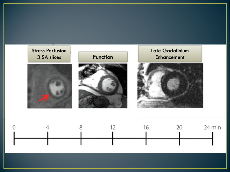
Fig.1: Assessment of myocardial ischemia with cardiac magnetic resonance imaging: the protocol
Stuff
Eleutherios Vidalakis, Cardiologist, specialised in cardiovascular imaging
MYOCARDIAL MAPPING WITH HEART MRI
A new, sensitive method of Covid 19-related myocardial damage detection
Mapping of the myocardium with a heart MRI is an advanced technique of multi-parameter mapping based on T1, T2 and T2* magnetic mapping sequences. This method provides us with high spatial analysis, so that we may be able to quantify lesions in every location within the myocardium. These new techniques are very specialized in detecting diffuse cardiac lesions, while assisting physicians, at the same time, to monitor the progress of myocardial disease, by comparing absolute numbers in successive measurements. Thus, myocardial mapping upgrades heart MRI diagnostic accuracy, by promoting this diagnostic method to reliability levels comparable to classic myocardial biopsies.
When is heart MRI myocardial mapping advisable?
- Myocarditis: diagnosis, monitoring and confirmation of full recovery. In 2018 this technique was officially incorporated in the myocardial diagnosis criteria.
- Myocardial attacks by autoimmune, rheumatic and systematic inflammatory diseases, such as lupus, rheumatoid arthritis, sarcoidosis, etc.
- Heart tumor characterizations
- HIV-related myocardial diseases
- Amyloidosis, Fabri’s Disease
- Differential hypertrophic myocardial conditions
- Coronary disease prognosis
- Covid 19-related heart lesions
MYOCARDITIS AND COVID-19
According to international medical studies, COVID-19 virus may attack the heart causing myocardial infarctions, or the most commonly known inflammatory myocardial condition, namely, myocarditis. Most importantly, myocardial inflammation is frequently non-symptomatic and subclinical, meaning that the patient has no way to know that the infection has affected his/her heart.
So, with just one examination we may investigate whether the coronavirus infection has affected the heart, even in patients with no cardiological symptoms. This fact gives us the opportunity to promptly intervene and protect the myocardium from permanent damage and subsequent symptoms in the future.
Post-Covid 19 patients now have the opportunity to examine their cardiac condition at Metropolitan General, with a specialized heart MRI protocol, based on myocardial mapping techniques, personally adjusted to each ex-Covid-19 patient by Dr. Eleftherios Vidalakis, our specialized Cardiologist and Cardiac Imaging Specialist.
Contact number: (+30)210 650 2510 - 11
Director
Eleftherios Vidalakis, Cardiologist specialized in cardiovascular imaging
Associate
Savvas Loizos, Cardiologist specialized in cardiovascular imaging
Opening hours: Monday-Friday 07:00 - 23:00 & Saturday 08:00 - 16:00
Coverage of emergencies 24 hours a day.
Contact Tel.: +30 210 650 2510 - +30 210 650 2511